
Imaging Anatomy of the Human Spine: A Comprehensive Atlas Including Adjacent Structures
Product details
| Management number | 231969626 | Release Date | 2026/06/18 | List Price | US$48.28 | Model Number | 231969626 | ||
|---|---|---|---|---|---|---|---|---|---|
| Category | |||||||||
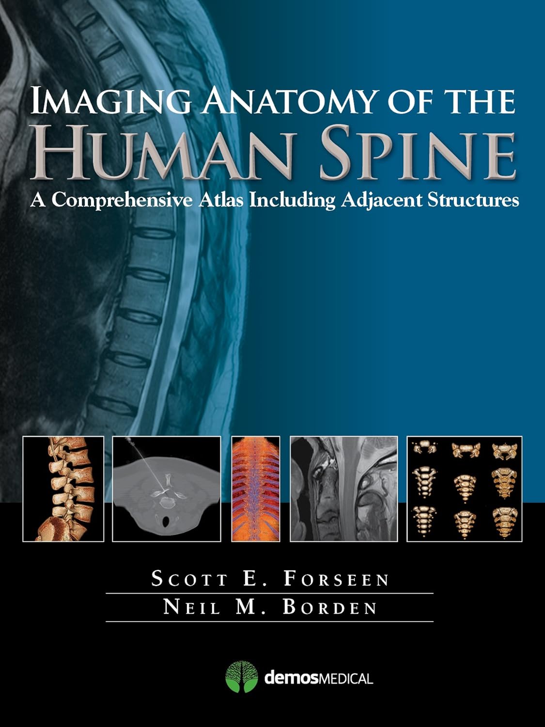
An Atlas for the 21st Century The most precise, cutting-edge images of normal spinal anatomy available today are the centerpiece of this spectacular atlasfor clinicians, trainees, and students in the neurologically-based medical specialties. Truly an ìatlas for the 21st century,î this comprehensive visual reference presents a detailed overview of spinal anatomy acquired through the use of multiple imaging modalities and advanced techniques that allow visualization of structures not possible with conventional MRI or CT. A series of unique full-color structural images derived from 3D models based on actual images in the book further enhances understanding of spinal anatomy and spatial relationships. Written by two neuroradiologists who are also prominent educators, the atlasbegins with a brief introduction to the development, organization, and function of the human spine. What follows is more than 650 meticulously presented and labelled images acquired with the full complement of standard and advanced modalities currently used to visualize the human spine and adjacent structuresóincluding x-ray, fluoroscopy, MRI, CT, CTA, MRA, digital subtraction angiography, and ultrasound of the neonatal spine. The vast array of data that these modes of imaging provide offer a wider window into the spine and allow the reader an unobstructed view of the anatomy presented to inform clinical decisions or enhance understanding of this complex region. Additionally, various anatomic structures can be viewed from modality to modality and from multiple planes. This state-of-the-art atlas elevates conventional anatomic spine topography to the cutting edge of technology. It will serve as an authoritative learning tool in the classroom, and as a crucial practical resource at the workstation or in the office or clinic.Key Features: Provides detailed views of anatomic structures within and around the human spine utilizing over 650 high quality images across a broad range of imaging modalities Contains several examples of the use of imaging anatomic landmarks in the performance of interventional spine procedures Contains extensively labeled images of all regions of the spine and adjacent areas that can be compared and contrasted across modalities Serves as an authoritative learning tool for students and trainees and practical reference for clinicians in multiple specialties Read more
| ASIN | B01BZ3010Y |
|---|---|
| XRay | Not Enabled |
| Format | Print Replica |
| ISBN13 | 978-1617051326 |
| Edition | 1st |
| Language | English |
| File size | 39.7 MB |
| Page Flip | Not Enabled |
| Publisher | Demos Medical |
| Word Wise | Not Enabled |
| Print length | 312 pages |
| Accessibility | Learn more |
| Publication date | December 17, 2015 |
| Enhanced typesetting | Not Enabled |
Bestseller ranking
Neuroscience


Correction of product information
If you notice any omissions or errors in the product information on this page, please use the correction request form below.
Correction Request Form